DIY 207 - 222 nm UV source for biologic death ray

Sorry to read about your condition and thanks for bringing up this matter. Quite an interesting read.

One thing i would actually worry about is over using this kind of technology though. If you grow up in a sterile environment you are likely to get sick with about anything if you wander into the ‘real world’… But of course it would be a big improvement for a lot of such applications. I hope it starts to get deployed in hospitals soon.

Do you think that your idea and implementation is something that makers of UVC packages for hospitals have not thought of?

Seems like companies such as www.americanultraviolet.com would have all of the answers and knowledge that you are seeking.

Yes they make UVC lamps for hospitals. But they are probably working around 254 nm. This is dangerous to people. My understanding is that 207 use is not yet approved by the FDA.

One problem with using light to sterilize is, it can’t get everywhere.

That is indeed true.

However, a significant factor in preventing surgical infection is the fact that the UVC will blow things out of the air.

In a surgical situation, most (if not all) of the tools and materials being used have been sterilized. More UV can’t hurt, and could improve the results. But a major element of contamination is airborne. Even with air filtration and sterilization protocols. Or a few contaminates from someone’s hair, nose, shoes, fingernails, breathing, and on and on. All these things can be a useful attack vector for UVC (207 nm).

The other application is to provide a localized source to protect the patient from pathogenic crap to be imported by nurses, family visits, food delivery persons, stethoscopes, the bed linen, water cups, blood pressure cuffs, IV insertions, not always really effectively washing hands, unfiltered air in the room, contagion of linen (e.g., would you like a blanket?), bed pans, just touching the bed or tray, and any other material effectively exposed to the surgical site. The potential contamination from just a few little tiny microbes can kill you. The personnel, equipment, the environment are simply not suitably disinfected.

Thinking how to create a completely sterile environment can hurt your head. But it is unlikely will you will figure out and block everything that is trying to kill the patient. And a hospital room simply cannot protect you.

You should consider 222nm lamps for a variety of reasons. We are working towards FDA approval for cornea disinfection.
Also show 99.99% air disinfection “kill-on-the-fly” single pass in an HVAC unit. UVC get’s less than 1%.
Good luck.

Just a note. As noted by Far-UV, I believe 222nm is going to be the target. I have not seen any useful semiconductor products at that frequency That certainly does not mean there are none. Or won’t be. The technology seems to be plasma discharge. This requires high voltage power supplies.

Far-UV. I am curious why the 222 nm radiation has to be approved for such a specific use. From what I have seen there is quite enough research to validate that the 222 nm radiation causes no harm to human surfaces. There are commercial devices outside the US. Why should it have to be approved for each human part in the US?

I am so excited about this technology. Here is a company claiming to have UVC LED capable of emitting any “single wavelength”. optical bypass filters can also be used to narrow wavelength to the desired frequency long as it emittes uv within the range one is looking for. The other factor to control is the intensity of the UVC.

Hi. I have been unable to get my computers to actually work for the last month and a half. One working. Go Microsoft; or simply be sent to Hell.

I am not sure if you are talking about another source or what I have posted. If you have new information; please post a link.

There have been multiple situations with hospitals hiding the fact that dangerous biologics are loose in hospitals (secure treatment areas, simple hospitalization and homes). Bad air is a significant component of spreading the diseases. No satisfactory methods are available for protecting patients, visitors, bed components, room contamination, nurses and other workers marching through the room touching everything. It goes on.

We are activity causing the propagation of human death so pigs and chickens can get big and inexpensive.

Was googling for affordable 222nm bulbs. Most are 254 nm and the search landed me at this forum.

Wan’t going to reply because I need to create a new account.
Then I realized that you did reply to a 2018 thread in 2020.
Anyway, I created the account so that I could reply with this link:

They have a 222nm uvc lamp, but I think their business is to sell expensive products, so it would be costly.

Hope it might be helpful, and hope your health is better now.
Still searching for 222nm products that I can DIY. The 254 nm ones are quite affordable, but I have to be out of the room to be safe.

I’m also interested in this, given recent events.

It seems like lasers and optical frequency doubling may be one way to do it. I’m not familiar with optics, need to spend more time learning.

I know this is an old thread, but I saw the discussion about Far UV-C lighting.
Here is a company who is starting to build these devices.

I just happened to send them an email earlier today with an inquiry into purchasing options.
I doubt that any products can be had immediately, but this was the most promising company I have seen for the Excimer bulbs which fit within the range you mentioned.
Good luck with your medical situation.

UV light is only line kill of sight except 185nm uv which also creates ozone as well as corona discharge units and ozone gas will find its way into every crevice. Ozone levels max are .8ppm and 5ppm is considered immediately dangerous So you can use ozone only if no living thing is is the room that can be harmed [includes plants] and you are willing to wait for the ozone levels to dissapate [1/2 life if 20 to 30 minutes]. an alternative is a Hydroxyl generator of OH generator which uses a UVC light with titantium dioxide coated filter or zinc coated mesh with moist air. Nanoparticle TiO2 and ZnO provide the greatest surface area and both metal are photocatalytic especially with humid air. UV light striking the metals produce OH ions aka Hydroxyl ions which float around in the air and destroy smoke and bacteria, etc. While there is some discussion that some VOCs can be broken down into equally hazardous byproducts, Hydroxyl generators are considered far safer than ozone generators and are used my many restoration companies that need to be working while the air is being decontaminated.

As far as the 207 to 222 nm uv being safe for human skin and eyes, this is true though counterintuitive since 222nm is a shorter wavelength than 254 uvc lamps so 222 would be more energetic and one would assume for it to be more damaging, but research has shown multiple times for Far uv of 297 to 222 to be safe. the researcher has suggested it might find use in surgeries to keep open wound from being infected by aerosol pathogens but not sure he has studied direct effects on tissue other than skin and eyes

You did not mention the company making these uvc leds? Most of the 60 watt uvc leds on amazon are fake, they just juse light blue leds to look like uvc but several utubes are out there with guys that have UV meters and these register no uvc no uvb or uva, too bad

While the use of Colloidal Silver as therapeutic medicine may be challenged as quack medicine [it is not going to cure your corona virus], It may well however kill the virus on surfaces since , the biocidal properties of Silver and many other metals is well researched, documented and proven. One can clean a pool or a pond with silver and copper electrodes When i was born, they put drops of Silver Nitrate into the eyes of every new born.

Below i pasted a researchgate article and i just read another comparing 9 metals for their biocidal properties but this one below used silver and silver worked better than all the other metals. One of the reasons utensils were made of Silver. Copper plated hospital surfaces often kill bacteria in 10 or 15 minutes and even the corona virus can not last longer than 3 or 4 hours on copper.rticlePDF Available

Germicidal Action of Some Metals/Metal Ions in Combating E. coli Bacteria in Relation to Their Electro-Chemical Properties

Citations (4)
References (67)
Figures (3)

Abstract and Figures

The germicidal properties of some metals and metal compounds were investigated in relation to their electro-chemical properties that may play a role in the inactivation of E. coli bacteria. These properties included the atomic and ionic radii, ionization energy, oxidation state, energy of formation with hydro-sulfide groups, and the redox potential of the metals. Cultures of E. coli bacteria with predetermined numbers of colony-forming units (CFU’s) were brought in con- tact with the metals as well as metal compounds, using Eosin methylene blue agar medium and sterilized, distilled water. The rate of inactivation was determined by counting the CFU’s at predefined intervals of time after inoculation. The experimental results showed that the rate of inactivation increases with increasing ionization energy of the metals. While the rate of inactivation increases with decreasing atomic radii for some of the transition metals, there is no ap- parent relationship between ionic radius and rate of inactivation for the metal compounds. In addition, non-transition group III metals such as aluminum and indium showed higher rates of in activation that are comparable to the action of silver. This is probably due to the increase in coagulation potential and the resulting adsorption of bacteria because of the larger number of ion is able electrons carried by these atoms. In general, there is a difference between the atoms and the ions in terms of their rate of inactivation. This difference increases amongst the transition metals that have lower oxidation potential, lower ionization potential as well as larger ionic radius. The results also showed that for the metals, adsorption through coagulation is an important fact or that is responsible for inactivation of E. coli. For the metal com- pounds, additional mechanisms such as direct reaction through complex formation, physico-chemical distortion of the cell structure through direct entry of the ions into the cell, may contribute towards greater inactivation.
Figures - uploaded by Ababu Teklemariam Tiruneh
Author content
Content may be subject to copyright.

Full-text (1)

Content uploaded by Ababu Teklemariam Tiruneh
Author content
Content may be subject to copyright.
Journal of Water Resource and Protection, 2013, 5, 1132-1143
Published Online December 2013 (http://www.scirp.org/journal/jwarp)
http://dx.doi.org/10.4236/jwarp.2013.512119
Open Access JWARP
Germicidal Action of Some Metals/Metal Ions in
Combating E. coli Bacteria in Relation to Their
Electro-Chemical Properties
Alakaparampil Joseph Varkey1, Mgidi Donald Dlamini1, Anaclet Bwampamye Mansuetus2,
Ababu Teklemariam Tiruneh3
1Department of Physics, University of Swaziland, Kwaluseni, Swaziland
2Department of Biological Sciences, University of Swaziland, Kwaluseni, Swaziland
3Department of Environmental Health Science, University of Swaziland, Mbabane, Swaziland
Email: varkey@uniswa.sz, mgidi@uniswa.sz, mansuetus@uniswa.sz, atiruneh@uniswa.sz
Received August 18, 2013; revised September 23, 2013; accepted October 21, 2013
Copyright © 2013 Alakaparampil Joseph Varkey et al. This is an open access article distributed under the Creative Commons Attribu-
tion License, which permits unrestricted use, distribution, and reproduction in any medium, provided the original work is properly cited.
ABSTRACT
The germicidal properties of some metals and metal compounds were investigated in relation to their electro-chemical
properties that may play a role in the inactivation of E. coli bacteria. These properties included the atomic and ionic
radii, ionization energy, oxidation state, energy of formation with hydro-sulfide groups, and the redox potential of the
metals. Cultures of E. coli bacteria with predetermined numbers of colony-forming units (CFU’s) were brought in con-
tact with the metals as well as metal compounds, using Eosin methylene blue agar medium and sterilized, distilled water.
The rate of inactivation was determined by counting the CFU’s at predefined intervals of time after inoculation. The
experimental results showed that the rate of inactivation increases with increasing ionization energy of the metals.
While the rate of inactivation increases with decreasing atomic radii for some of the transition metals, there is no ap-
parent relationship between ionic radius and rate of inactivation for the metal compounds. In addition, non-transition
group III metals such as aluminum and indium showed higher rates of inactivation that are comparable to the action of
silver. This is probably due to the increase in coagulation potential and the resulting adsorption of bacteria, because a
larger number of ions are able electrons carried by these atoms. In general, there is a difference between the atoms and
the ions in terms of their rate of inactivation. This difference increases amongst the transition metals that have lower
oxidation potential, lower ionization potential as well as larger ionic radius. The results also showed that for the metals,
adsorption through coagulation is an important fact or that is responsible for inactivation of E. coli. For the metal com-
pounds, additional mechanisms such as direct reaction through complex formation, physico-chemical distortion of the
cell structure through direct entry of the ions into the cell, may contribute towards greater inactivation.
Keywords: Metal Inactivation; Disinfection; Heavy Metal Toxicity; Water Treatment; Rate of Inactivation;
Escherichia coli
1. Introduction
Heavy metals are roughly delineated with a mass density
higher than 5 g/cm3. Out of the 90 elements found in na-
ture, 53 are heavy metals, constituting over 50% in abun-
dance [1]. By and large, the heavy metals belong to the
transition elements within complete d-orbital electrons.
They can easily lose electrons from the transition orbital
and form cations that have the ability to form complex
compounds with a reduction-oxidation potential [2,3].
Several of the heavy metals are important to biological
life. Among the 17 heavy metals that are useful to organ-
isms and the ecosystems, Fe, Mo and Mn are needed as
micronutrients [1]. On the other hand, Zn, Ni, Cu, V, Co,
W, and Cr do have importance as trace elements. How-
ever, they are toxic at high concentrations. The elements
As, Hg, Ag, Sb, Cd, Pb, and U do not have recognized
importance to biological life. Their toxicity, however,
has been reported [2,4,5].
1.1. Biocidal Properties of Heavy Metals
Silver ions are known from the long past to have a strong
biocidal effect against a wide range of pathogenic micro-
A. J. VARKEY ET AL.
Open Access JWARP
1133
organisms [6]. Copper has also been in wide use for me-
dical, cleaning, preservative and many other useful pur-
poses because of the low sensitivity of the human skin
and tissue to copper compared to the high level of sus-
ceptibility of microorganisms. The application of copper
for medicinal uses is quite ubiquitous [7]. However, cop-
per, at high concentration, shows toxicity to fish whereas
the level of toxicity of copper is greater in soft water than
in hard water.
Other heavy metals such as zinc and nickel do not rea-
dily undergo redox cycling like copper which is believed
to be responsible for its biocidal properties and is more
stable in its cationic oxidation states [8,9]. Therefore,
their use for medicinal application and as a biocide is li-
mited. However, zinc, while being an essential micronu-
trient to humans similar to copper, shows biocidal prop-
erties against fungi [10]. Zinc pyrithione is used as an an-
tifouling agent in paints [11]. Zinc solution at higher con-
centration is toxic to higher forms of life, including hu-
mans and its oxidative property at such concentration li-
mits its applicability as a biocide [12].
Contemporary applications of heavy metal inactivation
to disease causing viruses have been reported in medical
applications. The oxidation of the tyrosine phosphatase
in the cysteine of the Vaccinia H1 virus by copper ion led
to complete inactivation of the protein function of the vi-
rus [13]. On the other hand, the HIV-1 protease which is
useful for the replication of the virus has been neutralized
through exposure to a proportional concentration of cop-
per ions [14].
1.2. Mechanism of Inactivation of Pathogens by
Heavy Metals
A number of previous research works revealed several
mechanisms, by which the vital processes of the toxicity
of heavy metals to the cells are expressed. These
processes are discussed below.
1) Damage or destruction of the cell membrane:
The initial point of attack of heavy metals is under-
stood to be the plasma membrane [15-17]. Exposure of
microorganisms to higher concentrations of heavy metals
leads to deterioration in membrane structure leading, in
turn, to leakage of cellular solute materials such as potas-
sium and eventual death of microorganisms. Oxidation of
membrane lipids has also been reported to be the cause
of cell damage in higher organisms [17-19]. Not all hea-
vy metals are empowered, however, to cause damage
through cell destruction at a given concentration. Chan et
al. [20], for example, reported no significant effect by
other transition metals than copper which, when used
with 10 mM of H2O2, catalyzed peroxidation of rat eryth-
rocyte membrane lipid.
While extensive damage of the membrane integrity
leads to an obvious loss of cell function, even a small
change in the physico-chemical properties of cell mem-
branes can result in considerable alteration in the func-
tionalities of the several useful processes that are depen-
dent on the integrity of the cell membrane and which,
among others, includes the transport to protein activity
[21], phagocytosis [22], and ion permeability [21].
The bactericidal properties of several pathogens (Strep-
tococcus lactis, E. coli and P. aeruginosa), based on cell
membrane loss of functionality, have been reported to be
similar [23]. On the other hand, oxidation of susceptible
compounds within the cell structure requires a more ex-
tensive degradation of cells, possibly at higher concentra-
tion.
2) Formation of oxidative stress environment within
the cell structure.
The heavy metals in their ionic form (such as silver
ion, copper ion, cadmium ion, etc.) display an affinity for
binding with the so called sulfo hydroxyl group (SH
group) of the protein inside the cell [2]. For example, the
heavy metal ions that are completed with glutathione,
form a complex called bisglutathione. This complex fur-
ther reacts with oxygen molecules and results in the for-
mation of bisglutathionate, which is the oxidized form of
bisglutathione. The formation of the oxidized forms, bis-
glutathionate, is accompanied by the release of the com-
plexed metal ions and the formation of a reactive oxygen
species (such as hydrogen peroxide). The metal ions, now
freed from the complex, are able to react further in yet
another cycle for the formation of additional bisglutathi-
one and further oxidation [24,25].
3) Interference with vital functions of the cell.
The structure of a number of metal cation-based com-
pounds displays resemblance with metabolically useful
compounds. For example, chromate displays similarity
with sulfate, whereas arsenate shows similarity with phos-
phate. This means that the metal forms mentioned can
potentially interfere with the metabolic processes which
will need the important compounds such as sulfate and
phosphate [26].
4) Damage to DNA and protein through reduction of
metals.
A number of metal ions are able to acquire a reduced
state within the cell environment through processes in-
volving the cell enzymes or through other processes. For
example, chromium ion in the +6 oxidation state can be
reduced to the +3 state. Likewise, copper can be reduced
from the +2 oxidation state to the +1 state. The formation
of reduced metal cations is accompanied by the release of
reactive oxygen species which will oxidise and cause da-
mage or destruction to several of the cell components that
include the DNA and protein [27-29].
5) Displacement of Essential metals from their natural
binding sites.
Heavy metals, in general, have greater affinity binding
to the sulfohydroxyl groups than the essential metals.
A. J. VARKEY ET AL.
Open Access JWARP
1134
The complex formation with heavy metals as such occurs
through displacement of the essential metals or through
metal-ligand interaction.
1.2.1. Destruction of the Cell Barrier
The frontline attack of heavy metals begins with the cell
membrane. Heavy metals such as copper have been ob-
served attached to the plasma membrane of E. coli. In-
terestingly, steel that does not contain copper did not
attach to the cell membrane, indicating a selective me-
chanism by which heavy metals are attracted towards the
cell membrane [30]. The redox capability of heavy met-
als such as copper is observed as a factor in such selec-
tive binding.
The heavy metals that bind on the cell membranes
cause damage to the lipopolysaccharide on the outer plas-
ma membrane first. A significant alteration of the cell
permeability follows, leading to loss of the vital function
of the cell [16]. Cells rich with polyunsaturated fatty ac-
ids have been observed to lose much of the cell viability
[31]. A slight change in the structural property of the cell
membrane can induce a marked change in the activities
of several useful cell functions that depend on membrane
permeability barrier. These include the transport activi-
ties involving the proteins and the permeability of dif-
ferent ions [20].
1.2.2. Mechanism of Transportation of Heavy Metals
into the Cell Structure
The entry of heavy metals into the bacterial cell is done
through regulation involving the divalent cations or oxy-
anions [32]. However, heavy metals like other non-toxic
metals are indistinguishably transported to the cytoplasm
[2]. The heavy metals as such are not indistinguishable
from the other divalent metal ions or oxyanions that are
useful and pass across the membrane. These include
2
4
SO
,
2
4
HPO
, 2
Fe +,
2
Mg
+
,
2
Mn
+
, etc. Therefore,
subject to a certain concentration, heavy metals are af-
forded direct entry into the cell across the cell membrane.
However, toxicity effects result in a defense mechanism
that forms a barrier to the entry of heavy metals. This
phenomenon (metal resistance) will be discussed in a
later section of this paper. X-ray examination of the cell
structure has confirmed the existence of heavy metals
such as silver occupying electro dense environments
within the cell, caused by the entry of the heavy metals
[33]. Most of the observed biocidal actions against the
microorganisms, except those resulting from cell wall
damage, can only take place if the heavy metals are able
to enter the cell.
1.2.3. Inactivation, Damage and Destruction of the
Vital Contents of the Cell
Once the heavy metals find their way into the cell struc-
ture, they have an opportunity to form a complex reaction
with the protein structure inside the cell. The thiol groups
are known to be liable for the toxicity binding of heavy
metals [34]. Metals, as such, cause inactivation of the
protein inside the cells by reacting with the SH groups,
belonging to the protein structure [35]. Experimental stu-
dies with silver, indicating reaction of silver with thiol
groups, have been identified as critical for bacterial inac-
tivation [36].
Reactive oxygen species such as H2O2 are further
formed when oxygen reacts with metal-complexed SH
groups [24]. The reactive oxygen species so formed cause
oxidative stress within the cell, leading to inactivation of
the vital functions of the cell. The synergistic action of
oxidative stress and the metal complexed SH groups
causes a diminishing level of the activity of the vulner-
able enzymes [26].
A possible biocidal response of the stressed condition
within the cell is physiological changes involving the
DNA. The DNA is observed to shrink, leaving an elec-
tron sparse region in the cell while at the same time los-
ing its ability to replicate when shrunk. A dense electron
region is also formed towards the cell wall with a further
potential of reaction involving the heavy metals sur-
rounding the cell wall that can cause damage to the cell
wall [32]. The change of the conformal structure of nu-
cleic acid as well as cell proteins will result in loss of the
vital cell function, including loss of osmotic balance.
While normal interaction of metals, such as copper, is
possible with cell proteins of microorganisms, at elevated
concentrations the heavy metals may damage the protein
both on the outer cell membrane as well as inside [37,38].
Displacement of metals, interaction with the protein
binding sites leading to conformal changes to the protein
structure resulting in the inactivation of the protein vital
function have been reported at high heavy metal concen-
tration, notably silver and copper. These mechanisms can
be considered as direct attack [14,39]. Mediation by hea-
vy metals of proxy attack by free radicals also leads to
protein alteration and damage [40,41].
Extensive studies have been done on the damage, in-
activation and destruction of cell contents by copper ap-
plied as a biocide. Copper causes denaturing of the DNA
through helical structure disorders [42]. Several DNA
viruses revealed copper binding sites in every three nu-
cleotides [43]. A covalent binding of Cu2+ has been dem-
onstrated on DNA soaked with copper solution [44].
Multiple damage to nucleic acid is also caused by the
chain of redox reaction generated by the free radicals
[17]. The synergy between copper ion and the H2O2 oxi-
dant has been reported to be crucial in causing multiple
damage to nucleic acid. The absence of either of the two
may fail to cause the damage to the DNA [45]. Gunther
et al. [46] argue that copper plays a catalyst role in the
A. J. VARKEY ET AL.
Open Access JWARP
1135
conversion of H2O2 to free radicals. However, by way of
resistance and within the toxicity limits, stronger com-
peting ligands, such as glutathione and cysteine [47,48],
may remove copper away from the DNA in vivo. At high
heavy metal concentrations, inactivation of the protein’s
vital formulation has been reported due to displacement
of metals and interaction with the protein binding sites
that lead to conformal changes to the protein structures.
1.2.4. Complete Destruction
Silver, under an oxygenated environment, is capable of
forming reactive species and is as such known to achieve
a catalytic oxidation, leading to complete inactivation of
microorganisms. There is a synergy between reactive
oxygen species, such as hydrogen peroxide and silver,
which is assisting the inactivation process. The additive
action of reactive oxygen species and silver leads to com-
plete destruction of the cellular ingredients of microorga-
nisms and in their loss of vitality [49]. Denaturing of the
cell DNA by reactive oxygen species after the cell has
been subjected to chromium is well recorded [50,51].
The reactive oxygen species, in the case of chromium,
are reported to be produced through the reduction cycle
of chromium undergoing change from Cr (+6) state to Cr
(III) state.
Apart from destruction caused by oxidative stress in-
volving reactive oxygen species, physiological changes
within the cell structure are caused by the presence of
heavy metals inside the cell. The precipitation of the pro-
tein in the cell into a condensed state is caused by the
heavy metals. This physical (structural) change to DNA
and protein is, in addition to the chemical reaction taking
place between the heavy metal ions and complex com-
pounds, forming the cell contents [33]. The combination
of physico-chemical change taking place on the DNA
and protein, results in damage, inactivation and eventu-
ally death of the microorganisms [33].
1.2.5. Formation and Catalyzing the Action of
Reactive Oxygen Species
One process, by which reactive oxygen species, including
H2O2, are formed, is the interaction with oxygen of the
complex compounds formed by the reaction of heavy me-
tals with the non-specific thiol groups inside the cell [24].
Heavy metals such as silver, copper and cadmium pro-
duce oxidative stressed conditions within the cell by un-
dergoing complexation reaction with glutathione which,
in turn, results in oxidation reaction with molecular oxy-
gen and eventually the formation of reactive oxygen spe-
cies. The presence of increased levels of reactive oxygen
species results in an increased rate of peroxidation of
lipid, damage to the DNA and process changes to the cal-
cium and sulfhydryl homeostasis [17].
Studies surrounding the inhibitory action of chromate
suggested the presence of reactive oxygen species as a
result of the reduction of chromate [27-29]. The process
of chromate reduction from the Cr (VI) to Cr (III) passes
through Cr (V) intermediate first [52]. The process oc-
curs cyclically and produces overall high concentration
of reactive oxygen species, which will cause extensive
cell damage.
The redox cycling involving Cu2+ and Cu1+ is known
to generate reactive oxygen species in a manner shown
by the following reactions [17,53].
( ) ( )
22
Cu II O - Cu I O
+→ +
( ) ( )
22
Cu I H O Cu II OH OH
−−
+→ ++
The redox property is an important property of some
of the metals that are essential to the life of cells. How-
ever, this same property, as shown above, generates tox-
icity to the cell. The reactive oxygen species produced as
a result of such redox reactions are different forms of
oxygen-based compounds which include the super oxide
O2•-, the hydroxyl radical •OH, and hydroxyl anion OH.
Apart from metal-induced formation, the reactive oxygen
species are also formed by a number of natural sources,
including oxidative phosphorylation.
1.3. Subtle Differences and Grey Areas of the
Mechanism of Destruction
The actual mechanism of inactivation of pathogens by
heavy metals is not completely known and is still the
topic of active research. A considerable part of contem-
porary research involved observation of physiological
changes to cells which is increasingly leading to better
understanding of the mechanism of disinfection [33]. Of
all the metals, the biocidal mechanism of action of cop-
per has been well researched and is better understood.
Much of the explanation revolves around the redox capa-
bilities of copper [54].
There are, however, subtle differences in the types of
responses to oxidative stresses as a result of the presence
of different heavy metals. While the majority of the gen-
es give a response to the stress of oxidation of dismutase,
glutathione S-transferase, thioredoxin, and glutaredoxin,
the individual proteins affected by the changes that fol-
lowed differ from metal to metal. For example, cadmium
and chromium subject different sets of glutathione S-trans-
ferase to oxidation, suggesting that there are subtle varia-
tions of the heavy metal stresses and the physiological
state that follows [55].
The actual mechanism by which copper catalyses the
formation of free radicals is controversial. A study on E.
coli showed that copper did not catalyse DNA damage
that caused through oxidative stresses [47]. It is argued
that the majority of copper oxidative damage to the cell
A. J. VARKEY ET AL.
Open Access JWARP
1136
material may occur through Fenton mechanisms, particu-
larly at high concentrations [56]. Iron-mediated oxidative
damage by copper has been demonstrated [47].
The difference in biocidal properties between copper
and silver, on one side, and other closely related heavy
metals such as zinc and nickel, on the other, is not suffi-
ciently explained. One argument is the ability of copper
to undergo a cyclic redox reaction between Cu2+ and Cu+
that as a result, leads to damage of the cell lipid, protein
and DNA while metals with close properties such as zinc
and nickel do not go through such cyclic redox reactions
[8,9]. However, zinc still possesses some biocidal prop-
erties, such as being used as an anti-fungal agent for the
treatment of skin infections [10]. Zinc pyrithione is used
as an anti-fouling in paints [11].
The complexity of oxidative power of heavy metals
and their difference in power of oxidation is not suffi-
ciently explained in terms of the redox potential or the
redox states such as for copper and chromium. At high
concentrations, for example, zinc can promote oxidative
toxicity [57]. However, zinc, in ionic form, is danger-
ously toxic to higher organisms which limit its scope of
application [12]. While it can be argued that nickel also
possesses similar biocidal properties, it has a potential
risk in the forms of haematotoxic, immunotoxic, neuro-
toxic, genotoxic, nephrotoxic, hepatotoxic and carcinoge-
nic properties and is therefore not used extensively as
biocidal agent as much as copper is [58].
2. Materials and Methods
0.15 g of dried E. coli (source) was dissolved in 250 mL
of sterile distilled water. A 0.1 M solution of each of the
7 metal compounds was prepared using distilled water.
Metal plates 0.5 mm × 50 mm × 50 mm in size were pre-
pared. Eosin methylene blue agar was prepared and plac-
ed in a petri dish and incubated at 37˚C for a period of 8
hours to test its sterility 0.40 ml of the prepared E. coli
solution was put in two separate 250 ml glass beakers,
under a running Laminar Flow Hood.
The 0.5 mm × 50 mm × 50 mm metal was immersed
in one of the two glass beakers each containing 40 mL of
E. coli and in the other beaker 1 mL of the corresponding
compound was added, yielding a metal concentration of
2.5 mM. Both beakers were stirred. After every 5 min-
utes, 1 mL solution from each beaker was drawn and ino-
culated on the sterile media plate and then spread across
the plate using a spreader. The inoculated plates were in-
cubated at 37˚C for 48 hours. After this period, E. coli
colony counts were made using a digital colony counter.
The same procedure was applied for all the metal plates
and corresponding compounds. Concurrent with the tests
for each metal, a control blank test, without the metal or
compound, was run.
3. Results and Discussion
The experimental data consisting of the percentage of E.
coli colony forming units with respect to time were used
to study the kinetics of inactivation of E. coli by the vari-
ous metals and their ionic compounds employed. Figures
1 and 2 respectively show percentage of E. coli remain-
ing (in percentage units) against time of disinfection by
metals and their corresponding compounds. Both the
metals and their ionic compounds achieved complete in-
activation over a maximum period of 60 minutes. How-
ever, as can be seen from the graphs, the kinetics (rate of
inactivation) varies. Most of the metals used such as cop-
per, silver, cobalt and zinc are transition metals whose
properties make them suitable for inactivation. Some of
the properties of transitional metals include decreasing
reactivity because of smaller ionic radius, oxidizing pro-
perties, catalytic and complex formation properties. For
example, copper and silver are highly oxidizing, one of
the desirable properties for disinfection. Transition met-
als have smaller ionic radii that increase the ionization
energy and make them chemically less reactive. This pro-
perty, together with the ability of transition metals to
Figure 1. Percentage of E. coli remaining against time of in-
activation by the metals.
Figure 2. Percentage of E. coli remaining against time of in-
activation by metallic compounds.
A. J. VARKEY ET AL.
Open Access JWARP
1137
form complex compounds, might enable them to be bet-
ter available for complex formation with bacteria. An-
other property of the transition metals is their ability to
act as catalysts for oxidation of bacteria. The catalytic
properties of transition metals are achieved with their
variable valence, enabling them to form unstable inter-
mediate compounds. In other cases, the transition metals
provide suitable reaction surface compounds. The case of
copper undergoing a +1 and +2 redox cycle, while acting
as a catalyst for formation of reactive oxygen species has
been mentioned earlier.
The order of kinetics of disinfection of the transitional
metals, as shown in Figures 1 and 2, is explained by the
increasing rate of inactivation as one moves from left to
right across the periodic table for transition metals in the
order: titanium, cobalt, zinc, copper and silver. The oxi-
dizing property of zinc is less because the zinc group
(zinc, cadmium and mercury) have low boiling and melt-
ing points (even though their ionization energy is very
high) due to the low metallic bonding capabilities of the
s2 electrons. While zinc is electronegative, it is readily
oxidized into a stable form. Therefore, its oxidizing po-
tential is low. In addition, zinc has a low catalytic prop-
erty because the zinc structure (d10s2), indicates stability
of the d-shell with 10 electrons whereas copper (d10s1)
can have an oxidation state of 1 when the electron from
the s shell is removed, or an oxidation state of II or even
III when one or two electrons are taken from the d-shell.
Another property of the metals which is shared not
only by transitional metals, but also by group three(III)
metals used in the experiment (aluminum and indium), is
the coagulation potential. The poor shielding of the d-
orbital electrons of the transitional metals increases the
influence of the positive charge attraction of the nucleus
thus enabling the metals to act as coagulants against ne-
gatively charged bacteria. It is also known that coagula-
tion potential increases with increasing charge of the ion.
The higher charges of aluminum and indium play a grea-
ter role for coagulation. The results of the kinetics of the
inactivation experiment indicate that the group III metals
(aluminum and indium) as well as their ionic equivalents
achieved a faster rate of inactivation than most of the
transition metals.
Both aluminum and indium display similar kinetics of
inactivation, indicating that the property of inactivation is
mainly influenced by the charge of +3 which is common
to both ions of Group III metals in the periodic table.
This property of coagulation is especially important for
the metals because the electrostatic attraction of the met-
als can cause adsorption of bacteria onto metal surfaces
thereby facilitating further inactivation by coagulation,
hence making electrostatic attraction and adsorption an
important mechanism for inactivation of bacteria. Col-
loidal silver is commonly used for inactivation of bacte-
ria and coagulation is a useful property that can assist
inactivation in this regard.
3.1. Individual Metal/Ion Disinfection Kinetics
Evaluation
Figures 3 and 4 show the plots of percentage of E. coli
remaining against time of inactivation for the different
metals and the corresponding ionic compounds for com-
parison of the kinetics of inactivation of metal versus ion.
Aluminum and indium, which are the Group III metals,
have closer kinetics between the atoms and ions. Since
both have less oxidising properties, the coagulation (ad-
sorption) mechanism may have been similar for the at-
oms and ions. On the other hand, copper and silver also
show similar kinetics for the atom/ion inactivation. Both
copper and silver are known for their higher oxidation
potential and this mechanism may have been important
for both the atoms and the ions. By contrast, zinc, cobalt
and tin show a significant difference in the atom versu-
sion inactivation kinetics. The difference may be explain-
ed by the better inactivation properties of the ions through
reaction and complex formation because of the low oxi-
dation potential of these metals. These properties are not
much helpful for inactivation by the metals.
3.2. Relationship between E. coli Destruction and
Electro-Chemical Properties of the Metals
The variations in the rate of inactivation of the metals
and metal compounds with their ionization energy, which
is a measure of reactivity of the metals, are seen in Fig-
ures 5 and 6 respectively. There is a linear trenda de-
creasein the time required for inactivation with in-
creasing ionization energy. The regression coefficient of
the linear trend is 0.92 for the metals and 0.88 for the
metal compounds. The greater correlation of increasing
kinetics of inactivation with increasing ionization energy
for the metals, in comparison with the metal compounds,
might be an indication that for the metals, ions play a less
important role in the inactivation of E. coli. Silver and
titanium plot in Figure 6 as anomalies. The faster kinet-
ics of inactivation of silver may be explained partly by
the poorer shielding of the nuclear charge as the numbers
of d-orbital electrons increases.
Figures 7 and 8 show the trend of inactivation of E.
coli with respect to atomic and ionic sizes for metals and
their corresponding compounds, respectively. For the
metals, the trend of decreasing inactivation time with de-
creasing atomic size is apparent among the transition me-
tal series involving copper, cobalt and zinc. On the other
hand, the trend is weak for other metals such as alumi-
num, silver and indium. For the metal compounds, there
is an apparent decrease in inactivation time with increas-
ing ionic radius. However, the size of the ion may not
A. J. VARKEY ET AL.
Open Access JWARP
1138
(a) (b)
(c) (d)
(e) (f)
Figure 3. Percentage of E. coli remaining against time of inactivation for metals and their ionic counterparts (“Cls” refers to
“colonies”).
Figure 4. Percentage of E. coli remaining against time of
inactivation for silver and its ion counterpart (Clsrefers
to “colonies”).
Regression, R = 0.96
Figure 5. Variation of inactivation time by metals with ioni-
zation energy of metals.
A. J. VARKEY ET AL.
Open Access JWARP
1139
Regression, R = 0.88
Figure 6. Variation of inactivation time by metal compounds
with ionization energy of metals.
Figure 7. Variation of the time required for complete inac-
tivation by metals with atomic radius.
Figure 8. Variation of the time required for complete inac-
tivation by metal compounds with ionic radius.
be the only factor explaining inactivation. For example,
silver ion has a larger ionic size while its inactivation ki-
netics is fast, partly due to the poor shielding of the nu-
clear charge by the d-orbital electrons. The trend of faster
kinetics of inactivation might thus be more appropriately
linked to increasing nuclear charge. However, the combi-
nation of increasing nuclear charge and the comparative-
ly smaller ionic radii of transition metals still play a role
in the inactivation kinetics along with other properties of
the metals, such as higher oxidation potential, complex
forming ability, coagulation and catalytic action. These
are also related to the presence of easily transferable d
orbital electrons and due to poor shielding of the d orbi-
tals as the atomic number increases particularly for the
transition metals.
As explained in the preceding sections, heavy metals
generally react with proteins by combining with the thiol
groups that leads to the inactivation of the proteins. The
concentration of heavy metals used in the tests (2.5 mM)
may be high enough to cause membrane destruction and
protein denaturing. The data obtained in terms of the
time taken to achieve complete destruction of E. coli for
a number of metals has been compared with the free en-
ergy of formation of metal sulphides. Table 1 below shows
the solubility and free energy of formation of sulphides
for a number of metals.
A plot of the time taken to achieve complete inactiva-
tion of E. coli was plotted against the free energy of for-
mation of metal sulphides according to the data of Table
1. Figure 9 shows the result of such a plot. It is clear that,
generally, the time taken to achieve disinfection falls. As
expected, ions achieve faster inactivation than atoms.
However, for the metals with lower free energy of for-
mation (copper and silver) the difference between atom
and ion inactivation is considerably small compared with
metals of higher energy of formation. This partly indi-
cates the relative significance of formation of reactive
oxygen species compared to direct ionic oxidation of me-
tals, leading to DNA denaturing which is facilitated
through ion formation. On the other hand, atoms with
higher free energy of formation show a significant dif-
Table 1. Comparison of free energy of formation of metal
sulphides with time taken to achieve complete inactivation.
Compound Ksp
Ff0
(joules)
Time taken to
inactivate for
compound (min)
Time taken to
inactivate for
metals(min)
Ag2S 6.7 × 1050 9.3 15 25
CuS 6.3 × 1036 11.7 35 50
CoS 2.0 × 1025 19.8 50 60
ZnS 1.6 × 10²³ 47.4 45 60
25
Figure 9. Variation of inactivation time with free energy of
formation of heavy metals and metal compounds.
A. J. VARKEY ET AL.
Open Access JWARP
1140
ference in the rate of inactivation between atoms and ions.
The difference is expected to get wider when a lower
concentration of metals is used as is normally the case
with potable water disinfection with metals. On the other
hand, it is noticed from Figure 9 shows that the time
required for complete inactivation of E. coli falls much
faster at low free energy of formation. Therefore, al-
though cobalt and copper have a significant difference in
energy of formation, the time required for inactivation
does not vary much. The opposite is true for copper and
silver. Both of these have low free energy of formation
with a small difference in values (Table 1). However, the
time required for inactivation with silver is much smaller
than that of copper. This is a result of the increase in the
number of protons in the silver ions compared to copper
while the increased number of electrons is filled in the d-
orbitals with poor shielding of the protons’ attraction po-
tential.
3.3. Evaluation of Inactivation Kinetics with
Respect to Reduction-Oxidation
Properties Metals
Table 2 shows the standard redox potentials of the met-
als for E. coli in activation. The potentials are given with
respect to reduction of a hydrogen ion to a hydrogen gas
and show the relative potentials of the metals in reduc-
tion.
The high oxidation potential of silver and copper com-
pared to the other metals. On the other hand, metals on
the lower part of the table have limited oxidation poten-
tial as they are oxidized in turn. Zinc belongs to this set.
In order to examine the relationship between the ki-
netics of inactivation with the redox activity of the metals
which potentially influences the production of reactive
oxygen species that have oxidative power, the time taken
for complete inactivation of E. coli is tabulated against
standard redox potential of the metals as shown in Table
2. Figure 10 shows a plot of the time taken for complete
inactivation of E. coli against standard redox potential for
the metals used in the experiments. As expected, the ki-
netics is considerably faster for the atoms with higher
oxidation potential, such as copper and silver. By con-
trast, for zinc, aluminum and titanium, the oxidation po-
tential does not show a meaningful trend with the redox
potential. This observation is in line with previous ob-
servations that metals, such as zinc, show stability and do
not undergo redox cycling [12].
In fact, the metals aluminum and indium show an ano-
maly with respect to redox potential indicating that the
inactivation kinetics is not explained solely by oxidation
potential. Other factors such as coagulation should be ta-
ken into consideration.
Table 2. Redox potentials and time taken for complete inac-
tivation.
Atom Symbol Redox
potential
(mV)
Time taken to
inactivate for
compounds (min)
Time taken to
inactivate for
metals (min)
Silver Ag 0.8 15 25
Copper Cu 0.343550
Tin Sn 0.14 50 65
Cobalt Co 0.28 50 60
Indium In 0.342530
Zinc Zn 0.76 45 60
Titanium Ti 1.63 NA* 60
Aluminum
Al 1.66 35 40
*The metal compound for titanium was not available.
Figure 10. Variation of inactivation time with redox poten-
tial of heavy metals.
4. Conclusions
The rate of inactivation showed variation among the dif-
ferent metals and metal compounds. The rate of inactiva-
tion for several of the transition metals as well as the
Group III metals (aluminum and indium) showed a de-
creasing linear relationship with their respective ioniza-
tion energies. For the transition metals, the rate of inac-
tivation increases as one moves to the right along the row
of the transition series with the well-known property that
the ionic radii of the metals increase while doing so. In
addition, non-transition, Group III metals, such as alu-
minum and indium show faster inactivation comparable
to that of silver due probably to the increase in coagula-
tion potential and the resulting adsorption of bacteria as a
result of the larger charge carried by these atoms.
The experimental results showed that for the metals,
adsorption through coagulation seems to be the most im-
portant factor. Future experiments with metals aimed at
enhancing such a mechanism of inactivation might yield a
greater rate of inactivation than what is being realized at
present. For the metal compounds, additional mecha-
A. J. VARKEY ET AL.
Open Access JWARP
1141
nisms, such as direct reaction through complex formation,
physico-chemical distortion of the cells’ vital contents
through direct entry of the ions into the cell structure
may contribute towards greater inactivation. These fac-
tors are made evident through the association of the rate
of inactivation with redox potential and free energy of
formation of the metals with the sulfide groups. There is,
in general, a difference between the metals and metal
compounds in terms of their rate of inactivation. This
difference is larger for the transition metals that have
lower oxidation potential, higher ionization energy as well
as larger ionic radii.
The results also showed that the rate of inactivation for
the metals and metal compounds follow a trend with the
free energy of formation with sulfide, with the metals
having properties that enable them to form complexes
with SH groups within the cell as well as the redox po-
tential that enables the metals to catalyze the formation
of the cell-destroying reactive oxygen species. Metals
such as copper and silver, with higher oxidation potential
and higher free energy of formation, show a faster rate of
inactivation of E. coli. By contrast, the rate of inactiva-
tion of E. coli by metals with low redox potential and
free energy of formation with the sulfide groups is gen-
erally poor. The results point to a possible future work
with the active metals to increase their coagulation, reac-
tive and oxidation potential, with a view to enhancing the
inactivation of pathogens and increasing the scope of ap-
plication of heavy metals for disinfection purposes.
REFERENCES
[1] R. C. Weast, “CRC Handbook of Chemistry and Phys-
ics,” 64th Edition, CRC Press, Boca Raton, 1984.
[2] D. H. Nies, “Microbial Heavy-Metal Resistance,” Applied
Microbiology and Biotechnology, Vol. 51, No. 6, 1999,
pp. 730-750. http://dx.doi.org/10.1007/s002530051457
[3] M. Valls and V. de Lorenzo, Exploiting the Genetic and
Biochemical Capacities of Bacteria for the Remediation
of Heavy Metal Pollution,” FEMS Microbiological Re-
views, Vol. 26, No. 4, 2002, pp. 327-338.
[4] D. L. Godbold and A. Hüttermann, “Effect of Zinc, Cad-
mium and Mercury on Root Elongation of Piceaabies
(Karst.) Seedlings, and the Significance of These Metals
to Forest Die-Back,” Environmental Pollution, Vol. 38,
No. 4, 1985, pp. 375-381.
http://dx.doi.org/10.1016/0143-1471(85)90108-4
[5] C. W. Breckle, “Growth under Heavy Metals,” In: Y.
Waisel, A. Eshel and U. Kafkafi, Eds., Plant Roots: The
Hidden Half, Marcel Dekker, New York, 1991, pp. 351-
373.
[6] T. J. Berger, J. A. Sparado, S. E. Chapin and R. O. Bec-
ker, “Electrically Generated Silver Ions: Quantitative Ef-
fects on Bacterial and Mammalian Cells,” Anti Microb.
Agents, 1996, pp. 357-358.
[7] J. J. Hostynek and H. I. Maibach, “Hypersensitivity: Der-
matologic Aspects—An Overview,” Reviews on Environ-
mental Health, Vol. 153, No. 3, 2003, pp. 153-183.
[8] G. Borkow and J. Gabbay, “Copper as a Biocidal Tool,”
Current Medicinal Chemistry, Vol. 12, No. 18, 2005, pp.
2163-2175. http://dx.doi.org/10.2174/0929867054637617
[9] M. Valko, H. Morris and M. T. Cronin, “Metals, Toxicity
and Oxidative Stress,” Current Medicinal Chemistry, Vol.
12, No. 10, 2005, pp. 1161-1208.
http://dx.doi.org/10.2174/0929867053764635
[10] H. Rieth, “Killing of Pathogenic Fungi with Organic Zinc
Compounds. Fungicidal Aerosol Disinfection and Accu-
mulative Impregnation,” Mykosen, Vol. 11, No. 9, 1968,
pp. 667-670.
http://dx.doi.org/10.1111/j.1439-0507.1968.tb03410.x
[11] I. K. Konstantinou and A. T Albanis, “Worldwide Occur-
rence and Effects of Antifouling Paint Booster Biocides
in the Aquatic Environment: A Review,” Environment In-
ternational, Vol. 30, No. 2, 2004, pp. 235-248.
http://dx.doi.org/10.1016/S0160-4120(03)00176-4
[12] R. Eisler, “Zinc Hazard to Fish, Wildlife, and Invertebra-
tes: A Synoptic Review,Contaminant Hazard Reviews,
US Department of the Interior, Fish and Wildlife Service,
Laurel, 1993.
http://www.pwrc.usgs.gov/infobase/eisler/chr_26_zinc.pd
f
[13] J. H. Kim, H. Cho, S. E. Ryu and M. U. Choi, “Effects of
Metal Ions on the Activity of Protein Tyrosine Phospha-
tase VHR: Highly Potent and Reversible Oxidative Inac-
tivation by Cu2+ Ion,” Archives of Biochemistry and Bi-
ophysics, Vol. 382, No. 1, 2000, pp. 72-80.
http://dx.doi.org/10.1006/abbi.2000.1996
[14] A. R. Karlstrom and R. L. Levine, “Copper Inhibits the
Protease from Human Immunodeficiency Virus 1 by Both
Cysteine-Dependent and Cysteine-Independent Mecha-
nisms,” Proceedings of the National Academy of Sciences,
Vol. 88, No. 13, 1991, pp. 5552-5556.
http://dx.doi.org/10.1073/pnas.88.13.5552
[15] C. Cervantes and F. Gutierrez-Corona, “Copper Resis-
tance Mechanisms in Bacteria and Fungi,” FEMS Micro-
biology Reviews, Vol. 14, No. 2, 1994, pp. 121-137.
[16] Y. Ohsumi, K. Kitamoto and Y. Anraku, “Changes Induc-
ed in the Permeability Barrier of the Yeast Plasma Mem-
brane by Cupric Ion,” Journal of Bacteriology, Vol. 170,
No. 6, 1988, pp. 2676-2682.
[17] S. K. Stosh and D. Bagchi, “Oxidative Mechanisms in the
Toxicity of Metal Ions,” Free Radical Biology & Medi-
cine, Vol. 18, No. 2, 1995, pp. 321-336.
http://dx.doi.org/10.1016/0891-5849(94)00159-H
[18] P. R. Blackett, D. M. Lee, D. L. Donaldson, J. D. Fesmire,
W. Y. Chan, J. H. Holcombe and O. M. Rennert, “Studies
of Lipids, Lipoproteins, and Apolipoproteins in Menkes’
Disease,” Pediatric Research, Vol. 18, No. 9, 1984, pp.
864-870.
http://dx.doi.org/10.1203/00006450-198409000-00012
[19] A. H. Ding and P. C. Chan, “Singlet Oxygen in Copper-
Catalyzed Lipid Peroxidation in Erythrocyte Membran-
es,” Lipids, Vol. 19, No. 4, 1984, pp. 278-284.
[20] P. C. Chan, O. G. Peller and L. Kesner, “Copper (II)-Ca-
A. J. VARKEY ET AL.
Open Access JWARP
1142
talyzed Lipid Peroxidation in Liposomes and Erythrocyte
Membrane,” Lipids, Vol. 17, No. 5, 1982, pp. 331-337.
[21] J. R. Hazel and E. E. Williams, “The Role of Alterations
in Membrane Lipid Composition in Enabling Physiologi-
cal Adaptation of Organisms to Their Physical Environ-
ment,” Progress in Lipid Research, Vol. 29, No. 3, 1990,
pp. 167-227.
http://dx.doi.org/10.1016/0163-7827(90)90002-3
[22] S. V. Avery, J. L. Harwood and D. Lloyd, “Quantification
and Characterization of Phagocytosis in the Soil Amoeba
Acanthamoebacastellanii by Flow Cytometry,” Applied
and Environmental Microbiology, Vol. 61, No. 3, 1995,
pp. 1124-1132.
[23] H. Elzanowska, R. G. Wolcott, D. M. Hannum and J. K.
Hurst, “Bactericidal Properties of Hydrogen Peroxide and
Copper or Iron-Containing Complex Ions in Relation to
Leukocyte Function,” Free Radical Biology & Medicine,
Vol. 18, No. 3, 1995, pp. 437-449.
[24] A. V. Kachur, C. J. Koch and J. E. Biaglow, “Mechanism
of Copper-Catalyzed Oxidation of Glutathione,” Free Ra-
dical Research, Vol. 28, No. 3, 1998, pp. 259-269.
http://dx.doi.org/10.3109/10715769809069278
[25] D. H. Nies and N. Brown, “Two-Component Systems in
the Regulation of Heavy Metal Resistance,” In: S. Silver
and W. Walden, Eds., Metal Ions in Gene Regulation,
Chapman and Hall, London and New York, 1998, pp. 77-
103. http://dx.doi.org/10.1007/978-1-4615-5993-1_4
[26] P. Gong, O. Y. Ogra and S. Koizumi, “Inhibitory Effects
of Heavy Metals on Transcription Factor Sp1,” Industrial
Health, Vol. 38, No. 2, 2000, pp. 224-227.
http://dx.doi.org/10.2486/indhealth.38.224
[27] C. Cervantes, J. Campos-Garcia, S. Devars, F. Gutierrez-
Corona, H. Loza-Tavera, J. C. Torres-Guzman and R. Mo-
reno-Sanchez, “Interactions of Chromium with Microor-
ganisms and Plants,” FEMS Microbiology Reviews, Vol.
25, No. 3, 2001, pp. 335-347.
http://dx.doi.org/10.1111/j.1574-6976.2001.tb00581.x
[28] J. D. L. Singh, Carlisle, D. E. Pritchard and S. R. Patierno,
“Chromium-Induced Genotoxicity and Apoptosis: Rela-
tionship to Chromium Carcinogenesis,” Oncology Re-
ports, Vol. 5, No. 6, 1998, 1307-1318.
[29] Y. Suzuki, T. Sato, H. Isobe, T. Kogure and T. Murakami,
“Dehydration Processes of the Meta-Autunite Group Mi-
nerals, Meta-Autunite, Metasaléeite and Metatorbernite,”
American Mineralogist, Vol. 70, No. 8-9, 2005, pp. 1308-
1314. http://dx.doi.org/10.2138/am.2005.1568
[30] L. Nan, Y. Liu, M. Lu and K. Yang, “Study on Antibacte-
rial Mechanism of Copper-Bearing Austenitic Antibacte-
rial Stainless Steel by Atomic Force Microscopy,” Jour-
nal of Materials Science: Materials in Medicine, Vol. 19,
No. 9, 2008, pp. 3057-3062.
http://dx.doi.org/10.1007/s10856-008-3444-z
[31] S. V. Avery, N. G. Howlett and S. Radice, “Copper Toxi-
city towards Saccharomyces Cerevisiae: Dependence on
Plasma Membrane Fatty Acid Composition,” Applied and
Environmental Microbiology, Vol. 62, No. 11, 1996, pp.
3960-3966.
[32] M. Mergeay, D. Nies, H. G. Schlrgrl, J. Gerits and P.
Charles, Alcaligeneseutorophus CH34 Is a Facultative
Chemolithotroph with Plasmid-Bound Resistance to Hea-
vy Metals,” Journal of Bacteriology, Vol. 162, No. 1,
1985, pp. 328-334.
[33] Q. K. Feng, J. Wu, G. Q. Chen, F. Z. Cui, T. N. Kim and
J. O. Kim, “A Mechanistic Study of the Antibacterial Ef-
fect of Silver Ions on Escherichia coli and Staphylococ-
cus Aureus,” Journal of Biomedical Materials Research,
Vol. 52, No. 4, 2000, pp. 662-668.
[34] C. Rensing, B. Mitra and B. P. Rosen, “Insertional Inacti-
vation of dsbA Produces Sensitivity to Cadmium and
Zinc in Escherichia coli,” Journal of Bacteriology, Vol.
179, No. 8, 1997, pp. 2769-2771.
[35] A. L. Lehninger, D. L. Nelson and M. M. Cox, “Princi-
ples of Biochemistry,” 2nd Edition, New York, 1993.
[36] S. Y. Liau, D. C. Read, W. J. Pugh, J. R. Furr and A. D.
Russell, “Interaction of Silver-Nitrate with Readily Identi-
fiable Groups: Relationship to the Anti-Bacterial Action
of Silver Ions,” Letters in Applied Microbiology, Vol. 25,
No. 4, 1997, pp. 279-283.
http://dx.doi.org/10.1046/j.1472-765X.1997.00219.x
[37] F. Hussain, E. Sedlak and P. Wittung-Stafshede, “Role of
Copper in Folding and Stability of Cupredoxin-Like Cop-
per-Carrier Protein CopC,” Archives of Biochemistry and
Biophysics, Vol. 467, No. 1, 2007, pp. 58-66.
http://dx.doi.org/10.1016/j.abb.2007.08.014
[38] F. Hussain and P. Wittung-Stafshede, “Impact of Cofactor
on Stability of Bacterial (CopZ) and Human (Atox1)
Copper Chaperones,” Biochimica et Biophysica Acta, Vol.
1774, No. 10, 2007, pp. 1316-1322.
http://dx.doi.org/10.1016/j.bbapap.2007.07.020
[39] A. R. Karlstrom, B. D. Shames and R. L. Levine, “Reac-
tivity of Cysteine Residues in the Protease from Human
Immunodeficiency Virus: Identification of a Surface-Ex-
posed Region Which Affects Enzyme Function,” Archives
of Biochemistry and Biophysics, Vol. 304, No. 1, 1993, pp.
163-169. http://dx.doi.org/10.1006/abbi.1993.1334
[40] M. J. Davies, B. C. Gilbert and R. M. Haywood, “Radi-
cal-Induced Damage to Proteins: E.S.R. Spin-Trapping
Studies,” Free Radical Research, Vol. 15, No. 2, 1991, pp.
111-127. http://dx.doi.org/10.3109/10715769109049131
[41] R. T. Dean, S. P. Wolff and M. A. McElligott, “Histidine
and Proline are Important Sites of Free Radical Damage
to Proteins,” Free Radical Research, Vol. 7, No. 2, 1989,
pp. 97-103.
http://dx.doi.org/10.3109/10715768909087929
[42] R. B. Martin and Y. H. Mariam, “Metal Ions in Solution,”
Marcel Dekker, Basel, 2001.
[43] K. L. Sagripanti, M. L. Swicord and C. C. Davis, “Micro-
wave Effects on Plasmid DNA,” Radiation Research, Vol.
110, No. 2, 1987, pp. 219-231.
[44] B. H. Geierstanger, T. F. Kagawa, S. L. Chen, G. J. Quig-
ley and P. S. Ho, “Basespecific Binding of Copper (II) to
Z-DNA. The 1.3-A Single Crystal Structure of
d(m5CGUAm5CG) in the Presence of CuCl2,” The Jour-
nal of Biological Chemistry, Vol. 266, No. 30, 1991, pp.
20185-20191.
[45] E. Keyhani, F. Abdi-Oskouei, F. Attar and J. Keyhani,
“DNA Strand Breaks by Metal-Induced Oxygen Radicals
A. J. VARKEY ET AL.
Open Access JWARP
1143
in Purified Salmonella Typhimurium DNA,” Annals of
the New York Academy of Sciences, Vol. 1091, No. 1,
2006, pp. 52-64.
http://dx.doi.org/10.1196/annals.1378.054
[46] M. R. Gunther, P. M. Hanna, R. P. Mason and M. S. Co-
hen, “Hydroxyl Radical Formation from Cuprous Ion and
Hydrogen Peroxide: A Spin-Trapping Study,” Archives of
Biochemistry and Biophysics, Vol. 316, No. 1, 1995, pp.
515-522. http://dx.doi.org/10.1006/abbi.1995.1068
[47] L. Macomber, C. Rensing and J. A. Imlay, “Intracellular
Copper does not Catalyze the Formation of Oxidative
DNA Damage in Escherichia coli,” Journal of Bacteriol-
ogy, Vol. 189, No. 5, 2007, pp. 1616-1626.
http://dx.doi.org/10.1128/JB.01357-06
[48] A. Schrammel, D. Koesling, A. C. Gorren, M. Chevion,
K. Schmidt and B. Mayer, “Inhibition of Purified Soluble
Guanylyl Cyclase by Copper Ions,” Biochemical Pharma-
cology, Vol. 52, No. 7, 1996, pp. 1041-1045.
http://dx.doi.org/10.1016/0006-2952(96)00425-X
[49] R. Pedahzur, H. L. Shuval and S. Ulitzur, “Silver and
Hydrogen Peroxide as Potential Drinking Water Disin-
fectants: Their Bactericidal Effects and Possible Modes of
Action,” Water Science and Technology, Vol. 35, No.
11-12, 1997, pp. 87-93.
http://dx.doi.org/10.1016/S0273-1223(97)00240-0
[50] J. Aiyar, H. J. Berkovits, R. A. Floyd and K. E. Wetter-
hahn, “Reaction of Chromium (VI) with Hydrogen Pe-
roxide in the Presence of Glutathione: Reactive Interme-
diates and Resulting DNA Damage,” Chemical Research
Toxicology, Vol. 3, No. 6, 1990, pp. 595-603.
http://dx.doi.org/10.1021/tx00018a016
[51] K. E. Wetterhahn, J. W. Hamilton, J. Aiyar, K. M. Borges
and R. Floyd, “Mechanisms of Chromium(VI) Carcino-
genesis. Reactive Intermediates and Effect on Gene Ex-
pression,” Biological Trace Element Research, Vol. 21,
No. 1, 1989, pp. 405-411.
http://dx.doi.org/10.1007/BF02917282
[52] M. A. S. Fernandez, M. S. Santos, M. C. Alpoim, V. M.
C. Madeira and J. A. F. Vicente, “Chromium (VI) Inter-
action with Plant and Animal Mitochondrial Bioenerget-
ics: A Comparative Study,” Journal of Biochemical and
Molecular Toxicology, Vol. 16, No. 2, 2002, pp. 53-63.
http://dx.doi.org/10.1002/jbt.10025
[53] C. Barata, S. J. Markich, D. J. Baird, G. Taylor and A. M.
V. M. Soares, “Genetic Variability in Sub Lethal Toler-
ance to Mixtures of Cadmium and Zinc in Clones of
Daphnia magna Straus,” Aquatic Toxicology, Vol. 60, No.
1-2, 2002, pp. 85-99.
http://dx.doi.org/10.1016/S0166-445X(01)00275-2
[54] K. Apel and H. Hirt, “Reactive Oxygen Species: Metabo-
lism, Oxidative Stress, and Signal Transduction,” Annual
Review of Plant Biology, Vol. 55, No. 1, 2004, pp. 373-
399. http://dx.doi.org/10.1146/annurev.arplant.55.031903.
141701
[55] P. Hu, E. L. Brodie, Y. Suzuki, H. H. McAdams and G. L.
Andersen, “Whole-Genome Transcriptional Analysis of
Heavy Metal Stresses in Caulobactercrescentus,” Journal
of Bacteriology, Vol. 187, No. 24, 2005, pp. 8437-8449.
[56] C. Manzl, J. Enrich, H. Ebner, R. Dallinger and G.
Krumschnabel, “Copper-Induced Formation of Reactive
Oxygen Species Causes Cell Death and Disruption of Cal-
cium Homeostasis in Trout Hepatocytes,” Toxicology, Vol.
196, No. 1-2, 2004, pp. 57-64.
http://dx.doi.org/10.1016/j.tox.2003.11.001
[57] R. O. Wright and A. Baccarelli, “Metals and Neurotoxi-
cology,” The Journal of Nutrition, Vol. 137, No. 12, 2007,
pp. 2809-2813.
[58] K. K. Das, S. N. Das and S. A. Dhundasi, “Nickel, Its
Adverse Health Effects & Oxidative Stress,” The Indian
Journal of Medical Research, Vol. 128, No. 4, 2008, pp.
412-425.
... It is known that many metals by themselves (primarily silver and copper) also have antimicrobial properties (though the mechanisms of their antimicrobial activity are not yet known precisely [1]). Therefore, the deposition of metal films on fabrics is a promising method for creating antimicrobial coatings. ...
... Using the second system we were able first to clean the sample with Ar ions with an energy of 400…600 eV, then deposit of the metal films with help of metal ions with energy about 200 eV. Selection of titanium due to the fact that although the Ti itself does not exhibit antimicrobial activity [1], in the ambient air at the surface of the Ti film TiO 2 layer is fairly quickly formed, which already has antimicrobial properties. ...

... On the other hand, the antibacterial activity and mechanism of action have been gradually clarified that silver ions may cause Staphylococcus aureus (S. aureus) and Escherichia coli (E.coli) bacteria to reach an active but nonculturable (ABNC) state and eventually die, and also have been indicated to the mechanism of inactivation of pathogens by damages and destruction of the bacterial cell membrane. 4,5 The high antibacterial activity factor of Cu 2+ , Zn 2+ ions may be thought to be caused by binding bacterial surface proteins, cell membrane, and metal-binding complex formations. 6 However, bactericidal elucidation by metal-binding enzyme degradation due to inhibition of peptidoglycan (PGN) elongation and relationships between PGN synthesis and PGN hydrolase and autolysin has been still remained. ...
... Recent studies showing that non-indole-producing bacteria generate various oxygenases which may degrade indole or interfere with indole signaling [56,57]. Many oxygenase and reductase reactions may be involved in metal ion facilitation in bacteria [58]. e ability of S. oneidensis to reduce oxidized metals or nitrate e ectively has been identi ed as an important intrinsic activity of Shewanella species [59][60][61][62]. ...

... The presence of high concentrations of heavy metals can cause inactivation of pathogens through damage or destruction of the cell membrane, formation of oxidative stress environment within the cell structure, interference with vital function of the cell, damage to DNA and protein through reduction, and displacement of essential metals from their natural binding sites (Varkey et al., 2013). It is unlikely, however, that E. coli were affected by dissolved metals present in the treatment system pore water. ...

https://tpc.googlesyndication.com/safeframe/1-0-37/html/container.html
Article

Coli-aerogenes bacteria isolated from grass

March 2008 · Journal of Applied Microbiology
The numbers of coli-aerogenes bacteria on ungrazed herbage and on intensively grazed pasture were determined by inoculation into MacConkey's broth incubated at 30°, 37° and 44°. From the cultures producing acid and gas at 30° organisms were isolated on eosin methylene blue agar and classified according to the recommendations of the Coliform Sub-Committee. These micro-organisms were abundant in ... [Show full abstract]
Read more
Article

Microbial effects on colloidal agglomeration

Colloidal particles are known to enhance the transport of radioactive metals through soil and rock systems. This study was performed to determine if a soil microorganism, isolated from the surface samples collected at Yucca Mountain, NV, could affect the colloidal properties of day particles. The agglomeration of a Wyoming bentonite clay in a sterile uninoculated microbial growth medium was ... [Show full abstract]
Read more
Article

EFFECT O F C ROP R OTATIONS INVOLVING H OST A ND N ON-HOST P LANTS O N VESICULAR-ARBUSC ULAR M YCORR...

SUMMARY The effects of crop rotations involving two host (lettuce and lavender) and two non-host (cabbage and radish) plants on the development of mycorrhizal infection in host plants were studied in unsterile soil and in sterilized soil with or without rock phosphate and inoculated with Glomus mosseae. As in previous experiments, pre-cropping with non-host plants did not decrease VA infection in ... [Show full abstract]
Read more
Article
Full-text available

First Report of Xanthomonas gardneri Causing Bacterial Spot of Tomato in Ohio and Michigan

December 2011 · Plant Disease

In 2009 and 2010, outbreaks of bacterial spot characterized by significant fruit spotting occurred in at least 2,000 ha of commercial processing tomatoes in northwest Ohio and southeast Michigan. Losses were estimated at $7.8 million. Diseased fruit and foliage were collected from 32 Ohio and Michigan fields in 2010. Excised lesions from fruit and leaves were dipped briefly in 70% ethanol, air ... [Show full abstract]
View full-text
Article

Bioelectric Potentials of Tumor-Infected Plants

August 1953 · Plant physiology
The circuit for measuring potentials was essentially a bridge network using an FP54 electrometer tube (5) in vacuo with an input resistance of 1.16 x 10¹¹ ohms. With the voltages of the plants used the current was below 10~12 amperes. Contact with the plants was made by means of felt brushes moistened with a KCI solution flowing from test tubes into which were immersed Ag?AgCl electrodes. The ... [Show full abstract]
Read more

None of ushio care 22 uv bulbs are for sale, only oem manufacturers have access until the end of 2020 according to their website

Hi lightbringer,

Funny story, i once saw a man on the today show who had blue skin from drinking copious amounts of colloidal silver for a month or two. Apparently some sort of cyanotic reaction, not enough oxygen. That extreme case aside, While i would not expect Colloidal silver to knock out a Corona Virus infection, i personally have used Colloidal silver for sore throats and for mouth or gum infections or for rinsing wounds. Silver ions certainly have micro-biocidal activity, and this has been clearly demonstrated in the scientific literature. I posted one artice above and i will post another here. I myself use copper hydroxide (a fungicide in latex paint in the bottom of pots to stop root circling, copper sulfate is use to kill tree roots is sewer lines. I have use copper pipes as electrodes to kill algae in a pool. There are studies now on the superiority of copper plating over stainless steel for hospital surfaces. Nickel and Tin can also kill plant roots on contact

Metal‐based antimicrobial strategies - NCBI

www.ncbi.nlm.nih.gov › pmc › articles › PMC5609261
Jul 26, 2017 - A renewed interest in metals as antimicrobial and biocidal agents is ... to produce the metal nanomaterials with antimicrobial properties is a ...
by RJ Turner - ‎2017 - ‎Cited by 33 - ‎Related articles

Oligodynamic effect

From Wikipedia, the free encyclopedia
Jump to navigation Jump to search
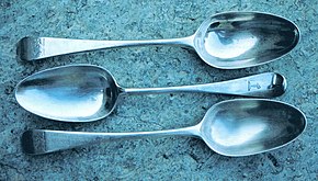
Silver spoons self-sanitize due to the oligodynamic effect

The oligodynamic effect (from Greek oligos "few", and dynamis "force") is a biocidal effect of metals, especially heavy metals, that occurs even in low concentrations.

In modern times, the effect was observed by Karl Wilhelm von Nägeli, although he did not identify the cause.[1] Scholarly texts from ancient India promoted the use of brass and silver in ritual cleansing practice as well as in consumption of food and drink. The ancient Indian medical text Sushruta Samhita promoted the use of specific metals in surgical procedures as a measure to prevent infection. Brass doorknobs and silverware both exhibit this effect to an extent.

Contents

+++++++++++++++++++++++++++++

It is now common knowledge that a variety of metal ions are toxic to bacteria (Nies, 1999; Harrison et al., 2004). Overall, the metals that are being increasingly considered for antimicrobial agents are typically within the transition metals of the d‐block, (V, Ti, Cr, Co, Ni, Cu, Zn, Tb, W, Ag, Cd, Au, Hg) and a few other metals and metalloids from groups 13–16 of the periodic table (Al, Ga, Ge, As, Se, Sn, Sb, Te, Pb and Bi). An interesting discovery made over 10 years ago that metals have strong efficacy against microbes growing as a biofilm (Teitzel and Parsek, 2003; Harrison et al., 2004). This was significant as a quintessential phenotype of biofilms is their antimicrobial resistance (Stewart and Costerton, 2001). Furthermore, metals have shown some efficacy on persister cells, the dormant variants of regular cells that were impervious to antibiotics (Harrison et al., 2005a,b).

Here is a nice picture

In this string, I don’t believe that anyone actually claims to have obtained 222 nm using an LED. At present the lowest wavelength available from an LED is 235 nm, and at this time that’s considered to be basic research rather than something available for consumer use. Anyone who does claim to be selling LED’s that emit in the range 207-222 nm is a fraud.